FIGURE 6.
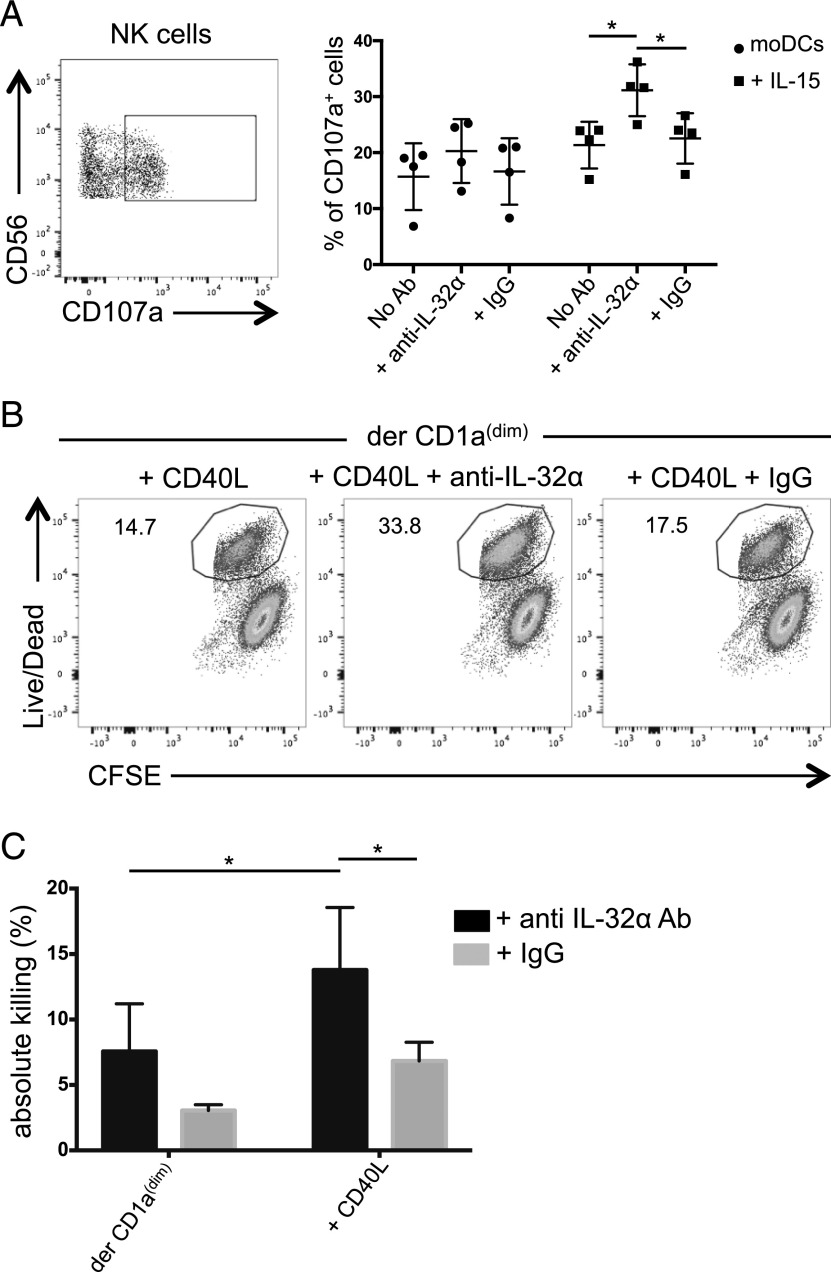
DC-derived IL-32α regulates NK cell–killing capacity. (A) moDCs were stimulated with IL-15 for 24 h or left unstimulated and cocultured with NK cells for another 24 h in the presence of anti–IL-32α mAb or an isotype-matched control mAb. Left, Plot shows the gating used to assess the percentage of CD107a+ NK cells. Right, Graph shows the percentage of NK cells that mobilized CD107a in response to a target cell line, n = 4. Values are calculated as average ± SD. (B) Sorted dermal CD1adim were stimulated overnight with CD40L (100 ng/ml) and cocultured with NK cells in the presence of anti–IL-32α neutralizing Ab or an isotype-matched control for 3 d. NK cells were then exposed to a target cell line. Dot plots show the percentage of dead target cells as indicated by the live/dead stain. (C) As in (B), the graph shows the percentage of dead target cells in the presence of anti–IL-32α Ab (black) or an isotype-matched control (gray). Values were normalized for five donors. Maximal killing was defined using IL-15–stimulated NK cell. *p ≤ 0.05.

