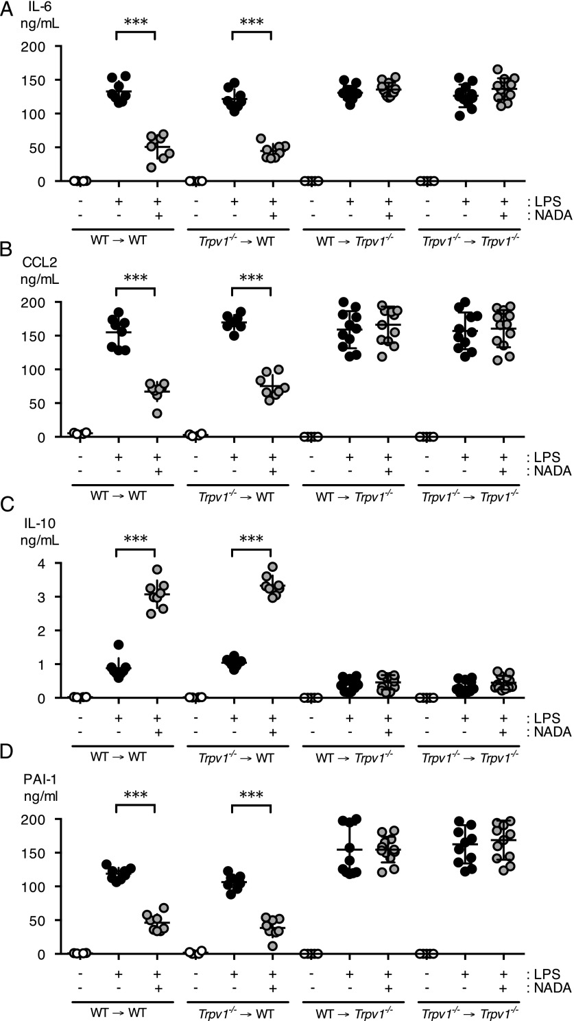
FIGURE 5.
Nonhematopoietic TRPV1 mediates the anti-inflammatory action of NADA in endotoxemic mice. (A–D) Irradiated CD45.1+ WT recipient mice were reconstituted with bone marrow from CD45.2+ Trpv1−/− mice (Trpv1−/− → WT). Irradiated CD45.2+ Trpv1−/− recipient mice were reconstituted with bone marrow from CD45.1+ WT mice (WT → Trpv1−/−) or Trpv1−/− mice (Trpv1−/− → Trpv1−/−). CD45.2+ WT mice were reconstituted with CD45.1+ donor bone marrow (WT → WT). Following engraftment bone marrow chimeras were challenged i.v. with LPS or vehicle and 5 min later were treated i.v. with NADA (10 mg/kg) or vehicle (n = 6–12 mice per group). Plasma levels of IL-6 (A), CCL2 (B), IL-10 (C), and PAI-1 (D) were quantified at 2 h. (A–D) NADA alone did not affect baseline levels of inflammatory mediators or PAI-1 in any of the bone marrow chimeras. The Mann–Whitney U test was used to calculate statistical significance. ***p < 0.001, LPS-treated mice in the presence or absence of NADA.

