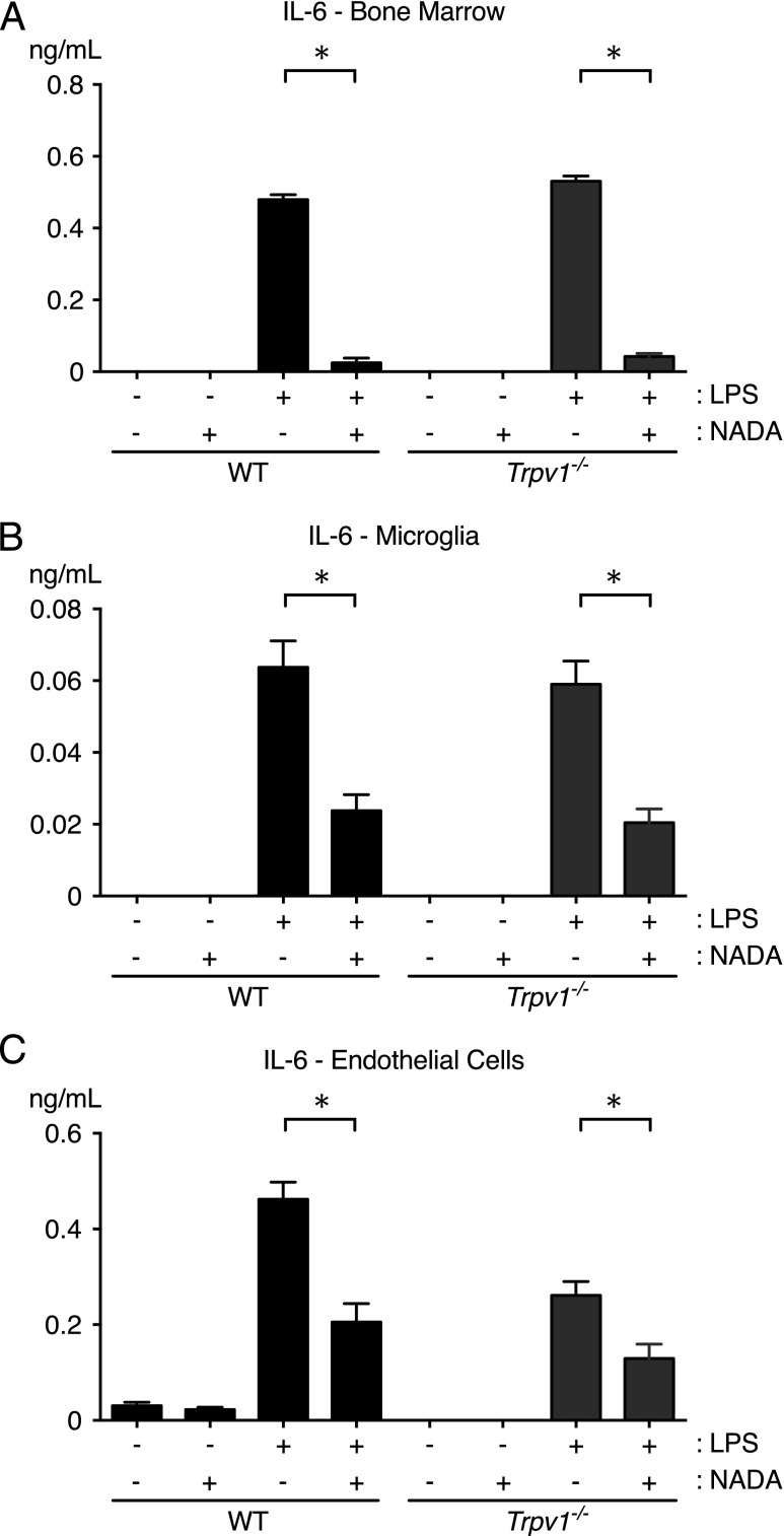
FIGURE 6.
TRPV1 does not mediate the anti-inflammatory activity of NADA in LPS-treated hematopoietic cells or endothelial cells ex vivo. Cultured primary murine bone marrow cells (A), microglia (B), and lung endothelial cells (C) were treated for 1 h with NADA (1 μM) and then stimulated with LPS (10 ng/ml) for an additional 6 h in the continued presence of NADA (n = 4 wells per group). IL-6 levels were quantified in culture supernatants. Identical results were observed with PBMCs and thioglycollate-elicited peritoneal macrophages (data not shown). *p < 0.05, LPS-treated cells in the presence or absence of NADA, Mann–Whitney U test.

