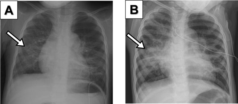
Figure 1. Comparative Chest Radiographs (CXRs) of a 2–year–old premature girl with HMPV infection.

(A) Initial CXR during admission to the hospital due to HMPV infection showing a focal infiltrate in the right lower lobe (arrow). (B) 48h later CXR shows progression of right lower lung opacities (arrow) that correlated with worsening of crackles, poor air entry, respiratory distress and hypoxemia.
