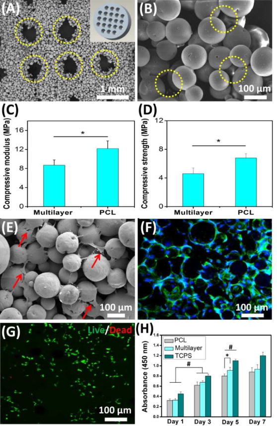
Fig. 2. Characterization and in vitro cellular evaluation of SLS-derived scaffolds.

(A) The multilayer scaffold presented a macroporous structure (yellow circles) corresponding to the designed model (inset in A). (B) SEM image at high magnification further showed that a large number of micropores (yellow circles) were generated among the microspheres. (C, D) Both compressive modulus and compressive strength of the multilayer scaffold were less than those of the PCL scaffold. (E) SEM image showed that cells attached to the multilayer scaffold (red arrows). (F) Confocal fluorescence image further showed that the attached cells exhibited a normal morphology (green: cytoskeleton, blue: nuclei). (G) Live/dead assay verified that the multilayer scaffold could support cell viability after 7 days of culture (green: live cells, red: dead cells). (H) The scaffolds could support cell expansion. Tissue culture polystyrene (TCPS) was used as a control. Data are shown as mean ± standard deviation for n = 5. (*) indicates a significant difference between groups (p < 0.05), (#) indicates a significant difference between day 1, day 3 and day 5 for the same group (p < 0.05).
