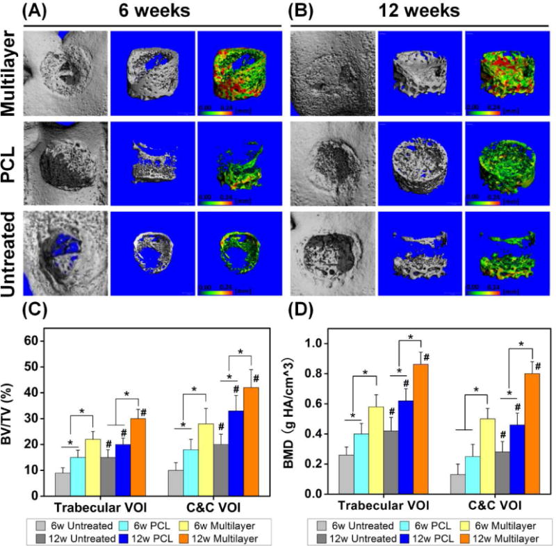
Fig. 4. Micro-CT analysis showed improved subchondral bone repair in multilayer scaffolds.

(A, B) The reconstructed images showed the joint surfaces (the first column) and the regenerated bone in the defect area (the second column) at 6 and 12 weeks post-surgery. The pseudo-color images (the third column) showed the thickness of new trabecular bone in the range 0–0.24 mm, as indicated by the color changing from green to red. (C, D) The quantitative micro-CT data further demonstrated that the multilayer group had the highest bone volume per total volume (BV/TV) and bone mineral density (BMD), in both trabecular volume of interest (VOI) and cartilage and cortical (C&C) VOI. BV/TV and BMD also increased for each group with time. Data are shown as mean ± standard deviation. (*) indicates a significant difference between groups (p < 0.05) and (#) indicates a significant difference between different time points for the same group (p < 0.05).
