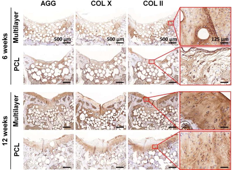
Fig. 7. Immunohistochemical staining for cartilage-specific proteins.

Aggrecan (AGG), collagen type X (COL X), and collagen type II (COL II) in sections from the multilayer and PCL groups were visualized at week 6 and week 12. Both AGG and COL II were abundant in the new cartilage of the multilayer group at week 12, and the expression of COL X was relatively low. The enlarged images of the selected region showed the distribution of COL II.
