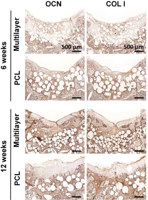
Fig. 8. Immunohistochemical staining for bone-specific proteins.

Osteocalcin (OCN) and collagen type I (COL I) in sections from the multilayer and PCL groups were visualized at week 6 and week 12. The subchondral bone region was enriched with OCN and COL I, and the staining for both proteins was remarkably stronger in the multilayer group than that in the PCL group.
