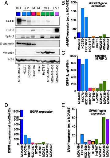
Fig. 1.

Characterization of TNBC subtype cell lines. a Lysates prepared from cells grown for 48 h in medium containing 10% FCS were evaluated for EGFR, HER2, SphK1, E-cadherin, and vimentin expression by western blot. Actin was measured as a loading control. The TNBC subtypes are basal-like 1 (BL1), basal-like 2 (BL2), immunomodulatory (IM), mesenchymal (M), mesenchymal stem-like (MSL), and luminal androgen receptor (LAR). Representative images from four similar experiments. b Relative IGFBP-3 mRNA levels averaged from duplicate cultures, normalized to MDA-MB-453 cells, analyzed by qPCR as described in Methods. c Secreted IGFBP-3 measured in duplicate by RIA specific for primate IGFBP-3 in 48-h conditioned medium (representative data from one of four similar experiments). d EGFR levels quantitated from the western blot in panel A, expressed relative to MDA-MB-453 cells. e Relative SphK1 mRNA levels averaged from duplicate cultures, normalized to MDA-MB-468 cells, analyzed by qPCR as described in Methods. EGFR epidermal growth factor receptor, HER2 human epidermal growth factor receptor-2, IGFBP-3 insulin-like growth factor binding protein-3, SphK1 sphingosine kinase 1
