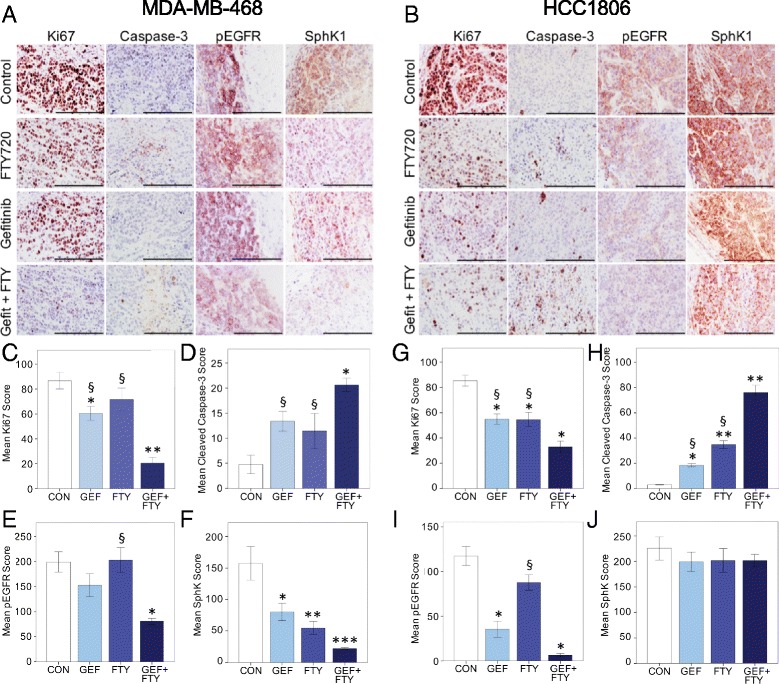
Fig. 6.

Immunohistochemical analysis of TNBC tumors from control, gefitinib-treated (GEF), FTY720-treated (FTY), and combination-treated mice. Tumors were stained for Ki67, cleaved caspase-3, pEGFR, and SphK1 as indicated. Scoring for each antigen was as described under Methods. a Representative staining of sections from MDA-MB-468 tumors. b Representative staining of sections from HCC1806 tumors. All summary data are mean values ± SEM, analyzed by one-way ANOVA followed by post hoc Tukey’s test. All ANOVA comparisons were highly significant for treatment except for SphK1 staining in HCC1806 tumors. Panels c-f MDA-MB-468 tumors. c Ki67 staining (n = 4–9 per group), * P < 0.05, ** P < 0.001 compared to control; § P < 0.001 compared to combination. d Cleaved caspase-3 staining (n = 4–10 per group), * P < 0.001 compared to control; § P = 0.05 compared to combination. e pEGFR staining (n = 3–5 per group), * P < 0.02 compared to control; § P < 0.02 compared to combination. f SphK1 staining (n = 3–5 per group): * P < 0.025, ** P = 0.005, *** P = 0.001 compared to control. Panels g-j HCC1806 tumors. g Ki67 staining (n = 10 per group), * P < 0.001 compared to control; § P = 0.01 compared to combination. h Cleaved caspase-3 staining (n = 8–10 per group), * P = 0.005, ** P < 0.001 compared to control; §P < 0.001 compared to combination. i pEGFR staining (n = 9–10 per group), * P < 0.001 compared to control; § P < 0.001 compared to combination. j SphK1 staining (n = 6 per group). Bar = 200 μm. pEGFR phosphorylated epidermal growth factor receptor, SphK1 sphingosine kinase 1
