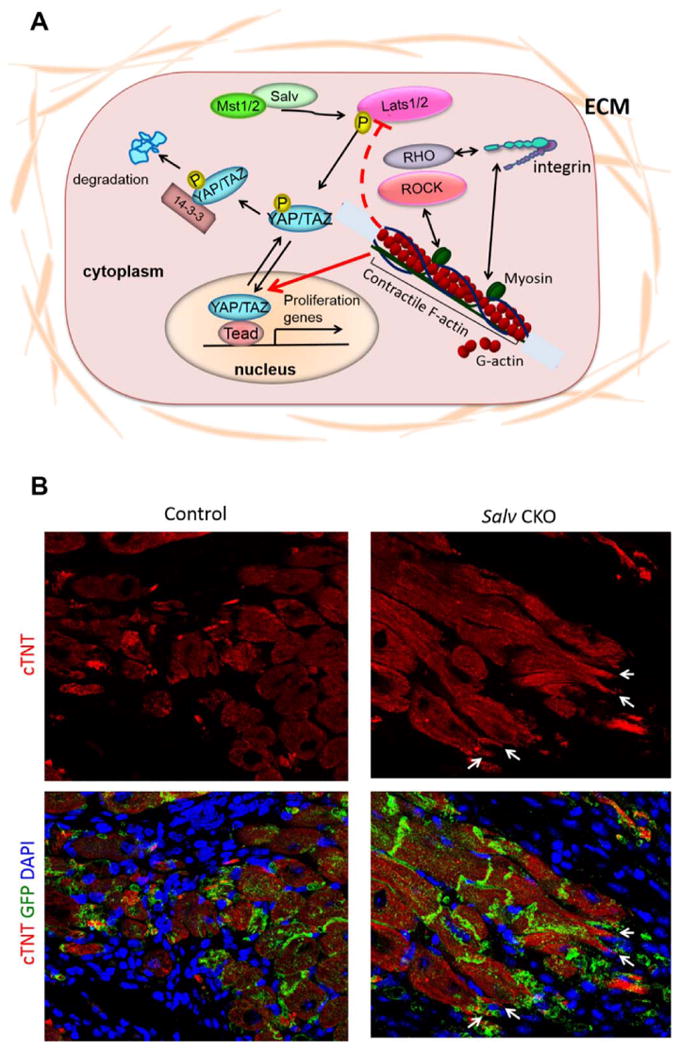
Figure 1. Hippo activity in adult cardiomyocytes.

A) Diagram of the Hippo pathway. The Hippo pathway is a kinase cascade composed of the core Mst and Lats kinases that regulate the phosphorylation status of downstream effectors Yap and Taz. Physiologic inputs that regulate Hippo pathway activity include signals from the extracellular matrix. The actin cytoskeleton also regulates Yap activity. Phospho-Yap is excluded from the nucleus and transcriptionally inactive. B) Phenotypes of Hippo deficient border zone cardiomyocytes. After experimental myocardial infarction in adult mouse hearts, Hippo-deficient (Salv CKO) cardiomyocytes extend sarcomere-filled protrusions. Control cardiomyocytes never make protrusions, suggesting that protrusion formation is required for efficient cardiomyocyte renewal. GFP marks the Myh6-expressing cardiomyocyte lineage.
