
Figure 3. Gram staining of colonic tissue.
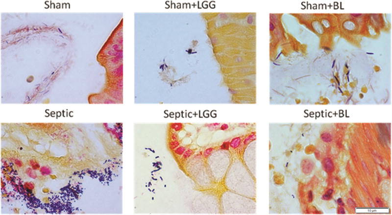
Increased numbers of Gram positive and Gram negative bacteria in the colon of septic mice compared to shams and mice treated with either Lactobacillus rhamnosus GG (LGG) or Bifidobacterium longum (BL).
Official websites use .gov
A
.gov website belongs to an official
government organization in the United States.
Secure .gov websites use HTTPS
A lock (
) or https:// means you've safely
connected to the .gov website. Share sensitive
information only on official, secure websites.
Increased numbers of Gram positive and Gram negative bacteria in the colon of septic mice compared to shams and mice treated with either Lactobacillus rhamnosus GG (LGG) or Bifidobacterium longum (BL).