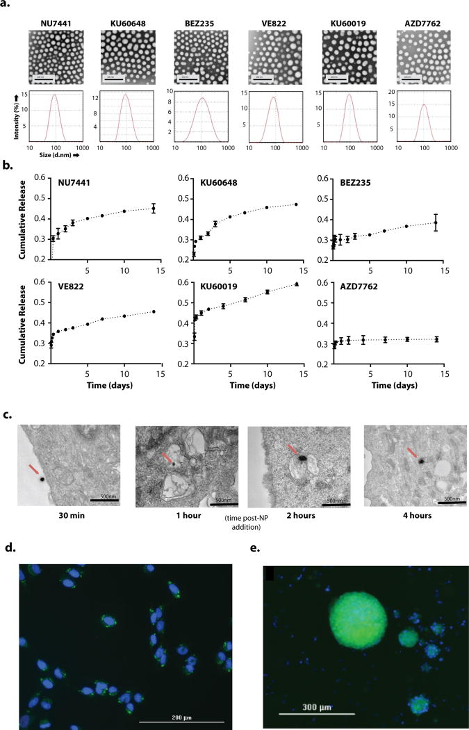
Figure 1. in vitro uptake characteristics and uptake of PLA-PEG NPs.
a. Transmission electron micrography (top row) and diffusion light scattering of hydrodynamic diameter (bottom row) of drug-encapsulated PLAPEG nanoparticles suspended in diH2O b. Release of encapsulated drugs from PLA-PEG NPs at 37°C suspended at 0.5mg NP/ml in 1×PBS spiked with 0.5% TWEEN80. c. in vitro transmission electron micrography imaging of cumarin6-loaded PLA-PEG NP uptake into U87 cells after incubation with PLA-PEG NPs suspended in cell culture media at 100mg/ml for 30 min, 1h, 2h, 4h. NPs indicated with red arrow. d., e. Fluorescence imaging of DAPI-stained SF188 cells (d.) and patient-derived DIPG spheroids (e.) after 4h incubation with coumarin6-loaded PLA-PEG NPs suspended in cell culture media at 100mg/ml. DAPI channel (nuclei) in blue, coumarin6 channel (NPs) in green.

