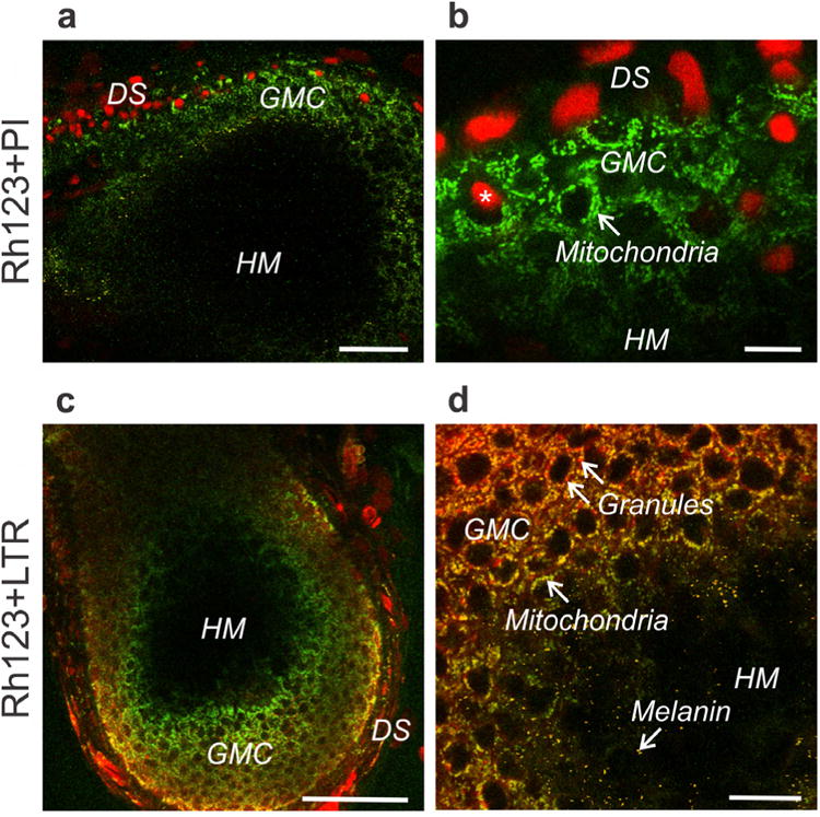
Fig. 2. Multiphoton imaging of cell viability, mitochondrial polarization and secretory vesicles in growing bovine hair follicles.

A,B. Bovine hair follicle loaded with green-fluorescing Rh123 and red-fluorescing PI and imaged by muliphoton microscopy. A scale bar = 50 μm. B scale bar = 25 μm. *, nonviable cell nuclei labeled with PI. C,D. A bovine hair follicle visualized with LTR (red, secretory vesicles) and Rh123 (green, mitochondrial ΔΨ). DS, dermal sheath; GMC, germative matrix cells; HM, hair matrix. C. scale bar =100 μm. D. scale bar = 25 μm.
