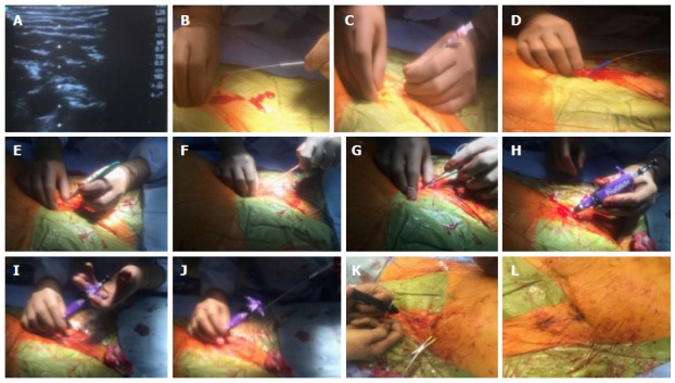
Figure 4.

Pre-close hemostasis technique during a transcatheter aortic valve replacement procedure. A-C: A calcium-free zone is visualized using real time ultrasound and access is obtained using a micropuncture needle system; D-G: The micropuncture system is exchanged for a 180 cm 0.035 wire and the skin tract is sequentially dilated with scalpel, 7 F sheath dilator and later a forceps; H: A Proglide is advanced over the wire into the arterial lumen, and return of pulsatile blood flow confirmed; I and J: After ensuring stable Proglide position, two sequential sutures are deployed at 10 and 2 0’clock; K and L: After the removal of transcatheter aortic valve replacement sheath and guide wire, pre-close sutures are sequentially locked to ensure hemostasis.
