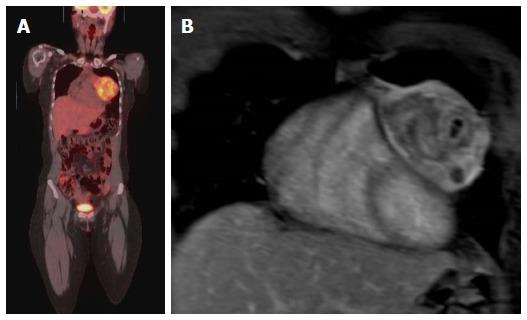
Figure 9.

Evaluation of the aggressiveness of the lesion and assessment of cardiac involvement. A: Evaluation of the aggressiveness of the lesion and assessment of cardiac involvement; Coronal PET-CT image in a patent with Ewing sarcoma of the left chest wall with direct compression of the left side of the heart; B: Coronal Post contrast T1-weighted image of the heart shows no evidence of cardiac invasion with clear separation of the mass from the heart, the mass was surgically removed and there was no evidence of cardiac invasion. PET: Positron emission tomography; CT: Computed tomography.
