Figure 2.
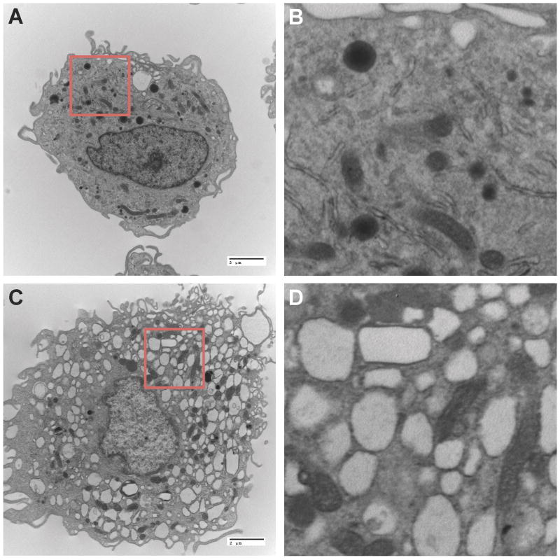
Macrophage uptake and storage of nanoformulated DTG prodrug crystals. A) Transmission electron microscopy (TEM) images of a human monocyte-derived macrophage (MDM) displaying eccentric nuclei, abundant cytoplasm, well-develop endoplasmic reticulum, lysosomes, and Golgi with intracytoplasmic vacuoles and a villus plasma membrane (magnified 6,500x). B) Higher magnification (30,333x) of highlighted area from panel A. C) Replicate human MDM treated for two hours with 100 μM nanoformulated DTG prodrug displays abundant intracellular vesicles within the cytoplasm rich with drug nanocrystals (magnified 6,500x). D) Higher magnification (30,333x) of highlighted region from panel C.

