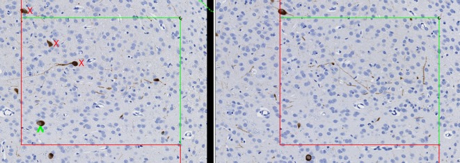
Fig. 2.
Application of the physical disector for estimation of cell or object number. Matching microscopic fields of view from serial sections are captured and placed side by side. A unique counting feature is chosen (in this case, the nucleus), and cells are counted if they are within the unbiased counting frame and the unique counting feature is present in one field but not the other. In this example, one cell is counted (green circumflex) because the nucleus is present in the field on the left but not in the field on the right. Other cells (red X) are not counted because the nucleus is not present on either side or the cell is outside of the counting frame. Rat brain, immunohistochemically stained for ChAT, 20× magnification.

