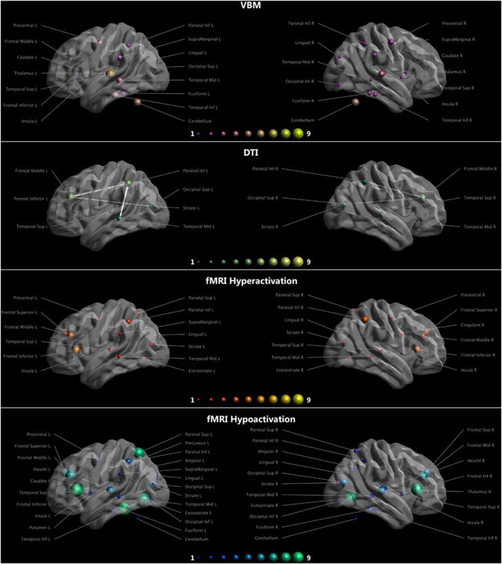
Figure 1.
Rows show the findings obtained with structural and functional MR techniques in DD subjects. The size and the color of the spheres reflect the amount of papers reporting differences in the specified area. Longitudinal fascicoli and arcuate fasciculus are shown as edges. fMRI findings are not divided by task. Task specific findings are available in Supplementary Tables 1 and 2. DD, developmental dyslexia; fMRI, functional magnetic resonance imaging. Figure was created with ExploreDTI (http://exploredti.com). DTI, diffusion tensor imaging; VBM, voxel-based morphometry.

