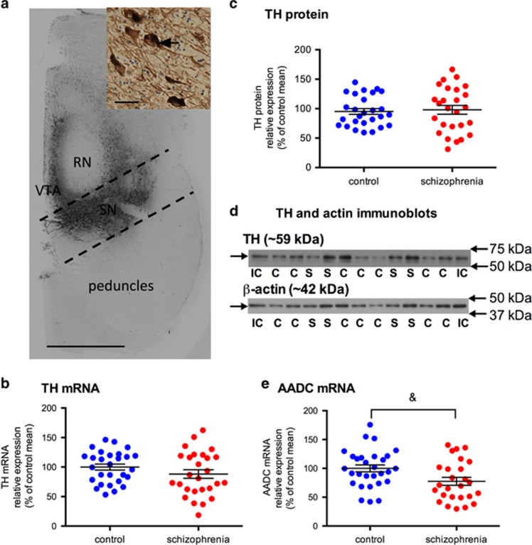
Figure 1.
Dopamine synthesis enzymes, TH mRNA and protein and AADC mRNA, levels in the substantia nigra in control (blue circles) and schizophrenia cases (red circles). (a) TH immunohistochemistry in a human midbrain representative of our cohort. Dark brown staining is TH expression in cell bodies and processes. Dashed lines bound the area of tissue dissected and homogenised to enrich for midbrain dopamine neurons of the substantia nigra (black). Scale bar=1 cm. Inset shows TH-positive neurons with TH expression in the cytoplasm (arrow) and processes, scale bar=100 μm. (b) TH mRNA in the substantia nigra was not significantly different between control and schizophrenia cases (F=0.74; df=54; P=0.395). (c) TH protein in the substantia nigra was not significantly different between control and schizophrenia cases (F=0.304; df=53; P=0.584). (d) A single TH protein band (~59 kDa) and a single β-actin band (~42 kDa) were detected in all samples using immunoblotting. (IC, internal control; C control; S, schizophrenia). (e) AADC mRNA was decreased 22.49% in the substantia nigra in schizophrenia cases when compared with controls; however, this only reached a trend level (F=3.417; df=54; P=0.070). Data are mean±s.e.m., &P<0.01. AADC, aromatic acid decarboxylase; RN, red nucleus; SN, substantia nigra; TH, tyrosine hydroxylase; VTA, ventral tegmental area.

