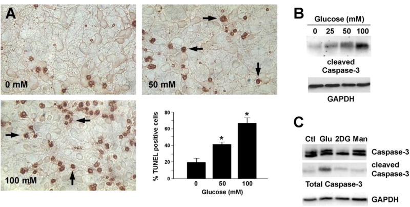
Fig 1. Glucose-mediated apoptosis of cortical neurons.
(A) E15 rat embryonic cortical neurons cultured in vitro for 6 d were treated with 0, 50 or 100 mM glucose for 24 h and apoptosis was measured using TUNEL staining. Arrows show the apoptotic cells. Glucose treatment significantly increased the number of TUNEL positive cells. p<0.05 by t-test. (B, C) Cortical neurons were treated with the indicated concentrations of glucose (Glu), 2-deoxyglucose (2DG, 100 mM) or mannitol (Man, 100 mM) for 24 h. Cell lysates were prepared in RIPA buffer and immunoblotted with an antibody against cleaved caspase-3 (B) or total caspase-3 (C). The blots were stripped and reprobed with an anti-GAPDH antibody. These experiments were repeated at least 3 times and a representative result is shown.

