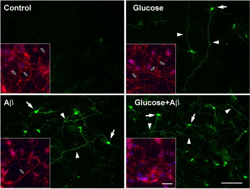
Fig 5. TauC3 immunostaining of rat embryonic cortical neurons.
E15 rat embryonic cortical neurons were grown on PLL-coated glass coverslips for 6 days and then treated with 100 mM glucose, 10 µM Aβ or both for 24 h. The cells were fixed with 2% PFA and stained for TauC3. Arrowheads indicate TauC3 staining along the neurites and arrows indicate staining in the cytoplasm. Insets: cells were stained with anti-neurofilament antibody (red) and DAPI (blue). Arrows in insets indicate the neurites. The bar represents 100 µm.

