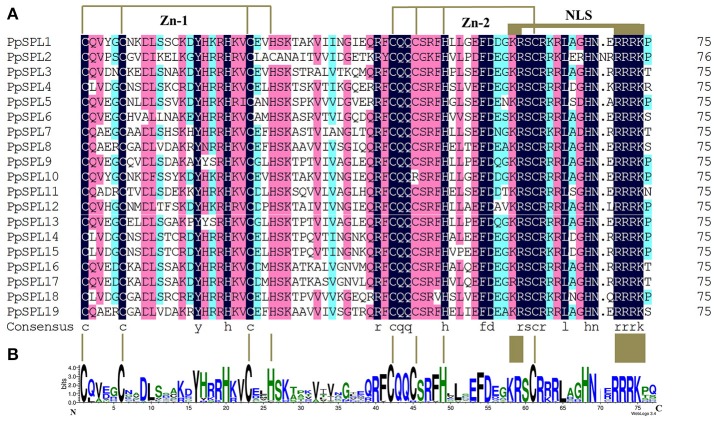
Figure 1.
Alignment of the SBP domains of PpSPL proteins. (A) Multiple SBP domain sequences from pear PpSPL proteins were aligned using DNAMAN software. Two conserved zinc finger structures (i.e., Zn-1 and Zn-2) and the nuclear localization signal are indicated. (B) Sequence logo of the SBP domain of PpSPL proteins. The overall height of each stack represents the degree of conservation at this position, while the height of the letters within each stack indicates the relative frequency of the corresponding amino acids.

