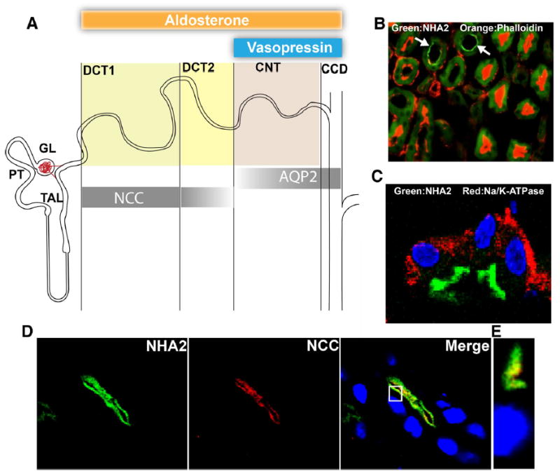
Fig. 1.

NHA2 is localized to the apical membranes of distal convoluted tubules in the mouse nephron. a Schematic representation of the segmentation of the mouse distal nephron and distribution of segment specific marker proteins. Shadings of bars indicate relative changes along the segments in immunohistochemical abundance. Target regions for aldosterone and vasopressin are shown on the top. Markers: NCC = Sodium chloride cotransporter, AQP2 = Aquaporin 2. b A merged microscopic image showing localization of NHA2 (green) and phalloidin (red) in tissue sections from mouse nephron. White arrows point to regions in tissue stained by NHA2 antibody. Green background observed in other areas is fixed-tissue auto-fluorescence. c A merged microscopic image showing subcellular distribution of NHA2 (green) and α-Na/K-ATPase (red) in tissue sections from mouse nephron. d Confocal microscopic image of a single tubule. Left panel NHA2 (green), middle panel DCT1 marker NCC (red), right panel merged image showing colocalization (yellow), nuclei are stained with DAPI (blue). e An orthogonal view of a slice from the confocal image (white box from the merged image in d)
