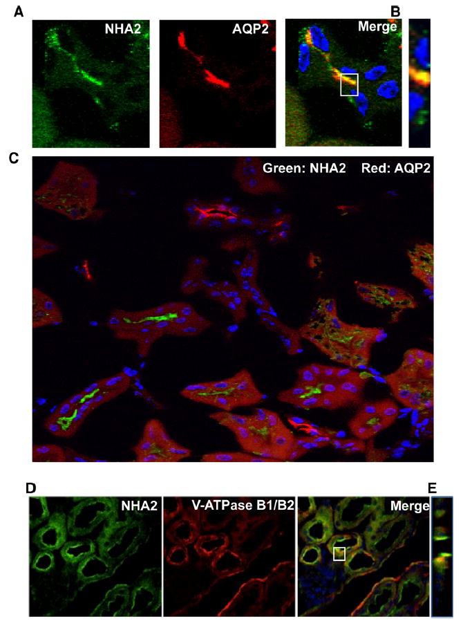
Fig. 2.

NHA2 partly localizes with Aquaporin 2 in the mouse nephron. a Confocal microscopic image of a single tubule. Left panel NHA2 (green), middle panel Aquaporin 2 (red), right panel merged image showing colocalization (yellow), nuclei are stained with DAPI (blue). b An orthogonal view of a slice from the confocal image (white box from the merged image in a). c A merged microscopic image showing NHA2 (green) and Aquaporin 2 (red) localized independently. d Microscopic images showing colocalization of NHA2 with V-ATPase in mouse nephron. Left panel indirect immunofluorescence of NHA2 (green), middle panel indirect immunofluorescence of V-ATPase B1/B2 (red), right panel merge of V-ATPase B1/B2 and NHA2 (yellow), Nuclei stained by DAPI (blue). e Orthogonal view of a slice from the merged confocal image (from d) showing colocalization of V-ATPase and NHA2
