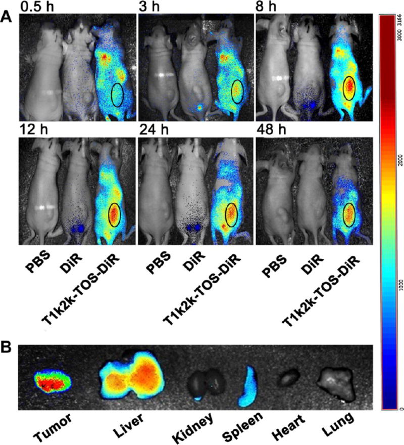
Figure 6.
(A) In vivo imaging of tumor-bearing mice after 0.5, 3, 8, 12, 24, and 48 h of administration with PBS, DiR, and T1k2k-TOS-DiR. The tumor area of the mouse treated with T1k2k-TOS-DiR is indicated by oval. (B) Ex vivo fluorescence imaging of excised organs: tumor, liver, kidney, spleen, heart, and lung after 48 h of treatment.

