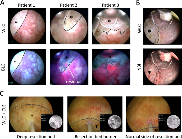
Fig.2.
Imaging of resection bed. (A) BLC can be used to detect residual tumor during TURBT as positive fluorescence is noted at the outside edges of the resection beds in these examples. There are residual tumors noted for patients 2 and 3 even within the resection bed on BLC that is not noted on WLC. (B) Residual tumor at the edge of the resection bed is noted on NBI. NBI images obtained with permission from [80]. (C) CLE can be used to interrogate the resection bed to determine adequate depth of resection. Features such as elastin strands, muscle fibers and perivesical fat can be visualized in the deep resection bed to verify adequate resection to the muscle layer in real time. All three WLC + CLE images with picture-in-picture are from the same resection bed. In the first image, elastin strands are noted on CLE in the deep resection bed. At the resection bed border, cautery artifact is noted with mostly absence of features seen in the muscle layer. On the normal side of the resection bed, CLE visualizes a capillary network typically seen in normal lamina propria. * – Indicates area within the resection bed. Dashed lines delineate the resection bed border.

