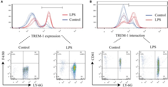
Figure 1.
FACS analysis of surface triggering receptor expressed on myeloid cells-1 (TREM-1) and its ligand on cells. Mice (n = 5) were inoculated with LPS for 6 h or mock treated with PBS as a control. The blood cells were collected, and red blood cells were removed for analysis of TREM-1 expression or analysis of the distribution of TREM-1-interacting proteins. All experiments were done in triplicate. (A) FACS analysis of TREM-1 expression on the cell surface with phycoerythrin (PE)-conjugated rat anti-mouse TREM-1 or PE Rat IgG2a, κ Isotype ctrl Antibody, allophycocyanin-conjugated anti-mouse F4/80, and Percp/cy5.5-conjugated anti-mouse Ly-6G. The signal for TREM-1 was specific (Figure S2 in Supplementary Material), and the cells expressing TREM-1 was further analyzed (Figure S3 in Supplementary Material). (B) FACS analysis of the distribution of TREM-1-interacting proteins on cells with Cy5.5-NHS-Ester-labeled recombinant extracellular domain of mouse TREM-1 (rTREM-1), PE/Cy7-conjugated anti-mouse CD41, and PE-conjugated anti-mouse Ly-6G. The cells expressing the ligand for TREM-1 were shown in details (Figure S4 in Supplementary Material).

