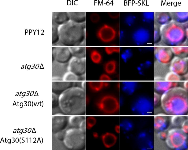
Fig. 2.
BFP–SKL localization during pexophagy. Strain PPY12 (wt) and the mutants were cultured in oleate medium to induce peroxisome and then shifted to nitrogen starvation medium (SD-N) to induce pexophagy. The pictures are taken after 2 h of starvation. The vacuoles are labeled red with FM4-64 and peroxisomes blue with BFP–SKL. Scale bars, 1 µm.

