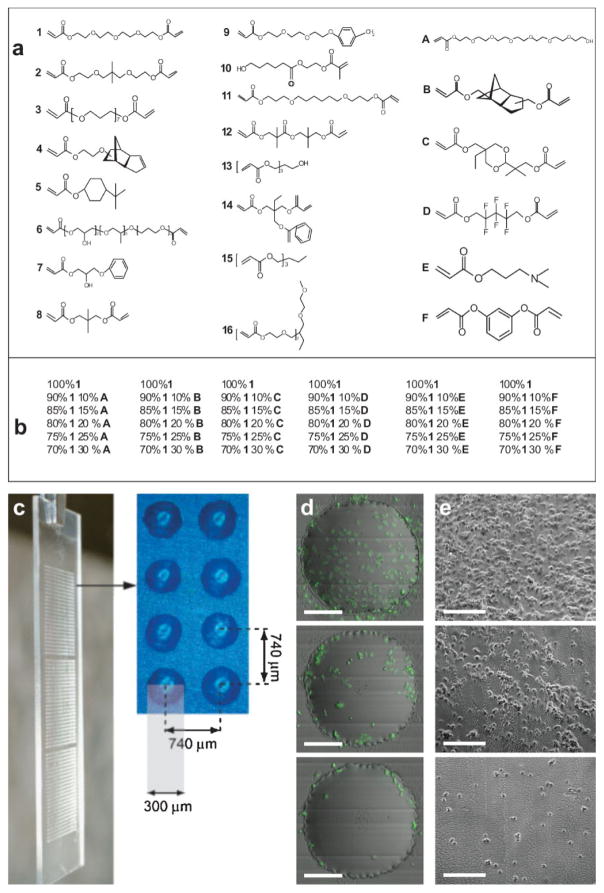
Figure 1.
Biomaterial array design: a) monomers used for array synthesis, b) 36 different combinations for the major monomer 1 with all six different minor monomers, c) photograph showing one polymer microarray in triplicate with eight polymer spots, to show dimension and separation. d) Merged images from fluorescence and scattered channel collected from iCys cytometry of three representative cell attachments (high, intermediate, low) on the polymer spots. The scale bar in the figure is 100 μm. e) Reproduced cell attachment on large-scale polymer films; the scale bar in the figure is 200 μm.

