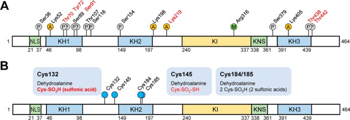
Figure 3.
Schematic diagram of hnRNP K domains and its PTMs. hnRNP K protein spots in silver-stained gels (Fig. 1C) were analyzed for PTMs using nanoUPLC-ESI-q-TOF MS/MS. A, all of the identified sites for phosphorylation (P circles), acetylation (A circles), and methylation (M circle) are indicated. PTMs of residues in red are disappeared after heat shock treatment (45 °C, 30 min) following 4 h of recovery. B, various identified Cys modifications are indicated. PTMs in red disappeared after heat shock treatment (45 °C, 30 min) following 4 h of recovery. PTM in bold is spot 2– and 4–specific (Fig. 2A). NLS, nuclear-localization signal; KH, K homology; KI, K-protein-interactive; KNS, nuclear shuttling domain.

