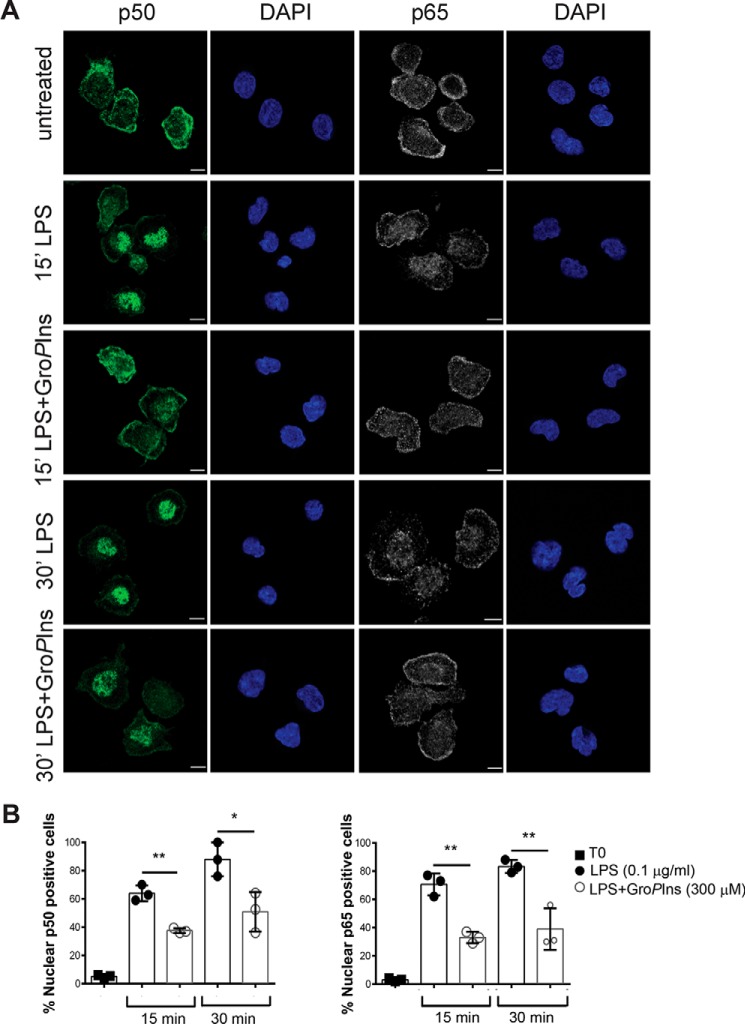
Figure 4.
GroPIns reduces the LPS-induced nuclear translocation of NF-κB. Representative confocal microscopy images of NF-κB intracellular localization in human peripheral blood monocytes are shown. A, cells were incubated at 37 °C for 20 min in the presence or absence of 300 μm GroPIns and then treated for 15 and 30 min (′) with 0.1 μg/ml LPS. Cells were then fixed and processed for immunofluorescence (see “Experimental procedures”). The intracellular localization of NF-κB was detected using antibodies specific for p50 and p65 proteins revealed with Alexa Fluor 488-labeled (green) and Alexa Fluor 568-lebeled (gray) secondary antibodies, respectively. Nuclei were stained with DAPI (blue). The samples were analyzed on a laser scanning confocal microscope (LSM710) equipped with a 63× objective. B, the nuclear translocation of NF-κΒ on cells left untreated or treated with LPS in the presence or absence of GroPIns has been quantified by randomly counting 100 cells/sample and expressing the percentage of cells with nuclear staining of p50 or p65. Quantification of nuclear events is presented as the mean ± S.D. of three independent experiments. Statistical analysis was performed using Student's t test (*, p < 0.05; **, p < 0.01). Error bars represent S.D. Scale bars, 5 μm.

