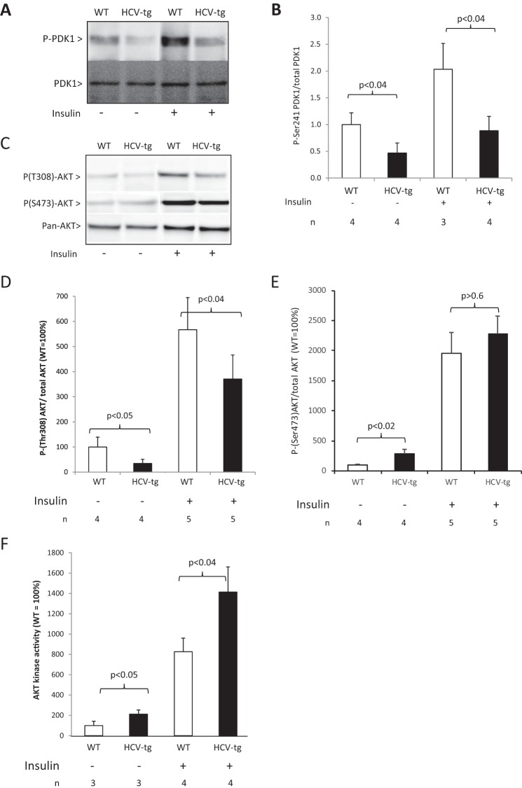
Figure 6.
A, Ser(P)241-PDK1 assessed by Western blotting in liver extracts from WT and HCV-tg mice injected with insulin or vehicle. The results were normalized using total PDK1 expression (representative animals). B, quantitative results for Ser(P)241-PDK1 protein levels. C, Thr(P)308-Akt assessed by Western blotting in liver extracts from WT and HCV-tg mice injected with insulin or vehicle. The results were normalized using total Akt expression (representative animals). D, quantitative results for Thr(P)308-Akt protein levels. E, quantitative results for Ser(P)473-Akt protein levels. F, analysis of Akt activity by kinase assay in hepatic lysates from WT and HCV-tg animals. Akt activity was normalized to the relative expression of Akt in the lysates and is expressed as the percentage of WT Akt activity.

