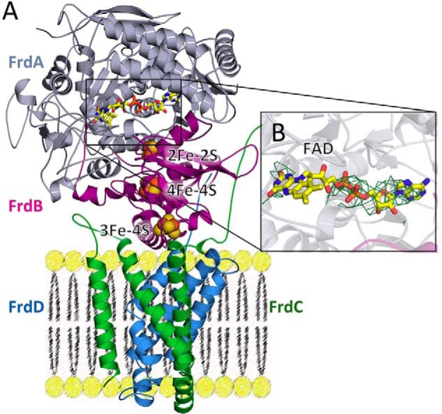
Figure 5.

Structure of QFR containing the FrdAE245Q mutation. A, structure of QFR-FrdAE245Q. The flavoprotein (FrdA) is shown in gray, the iron-sulfur protein (FrdB) is shown in magenta, and the membrane subunits (FrdC and FrdD) are shown in green and blue, respectively. The locations of the cofactors (FAD as yellow sticks, and iron-sulfur clusters as spheres) are also shown. B, |Fo| − |Fc| electron density maps contoured at 3σ were calculated in phenix.refine after the removal of FAD from the input PDB file. The quality of the electron density is consistent with the resolution for the adenine dinucleotide, but there is no interpretable density for the isoalloxazine ring.
