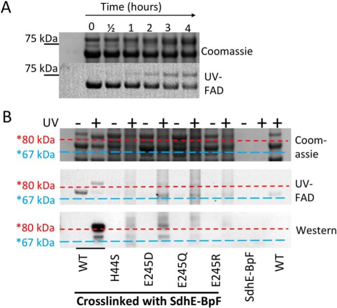
Figure 7.

Cross-linking between wild-type and variant FrdA subunits and SdhE-R8BpF. Purified SdhE-R8BpF was mixed with lysate containing FrdA. Following UV exposure, samples were immobilized on Ni2+ resin, washed, and eluted with SDS-PAGE loading buffer prior to separation on an SDS-PAGE gel. A, time course of cross-linking of SdhE-R8BpF with wild-type flavoprotein over 4 h after exposure to UV light. The position of the 75-kDa molecular mass marker is highlighted. B, comparison of flavin fluorescence for detection of covalent FAD, Coomassie-stained SDS-PAGE and anti-His6 Western blot analysis of cross-linked samples. Horizontal lines were drawn after aligning the Coomassie, UV, and Western blot analyses to assist with identifying the molecular weights of the bands. The blue dashed lines are drawn just under the location of the FrdA subunit (∼67 kDa) and red dashed lines are drawn just under the approximate location of the FrdA-SdhE complex (∼80 kDa).
