
Figure 5.
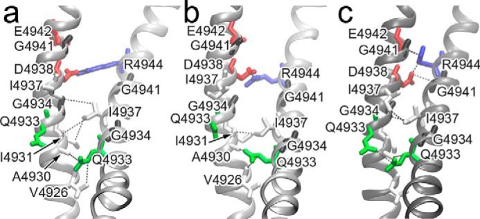
Intersubunit contacts for the S6 helix. Contacts are shown for the RyR1 open-channel model (a), the RyR1 open-channel structure (PDB code 5TAL) (b), and the RyR1 closed-channel structure (PDB code 3J8H) (c). The contacting residues are shown in stick representation and colored by residue type: red, acidic; blue, basic; green, polar; and white, aliphatic. The two S6 glycines are colored black.