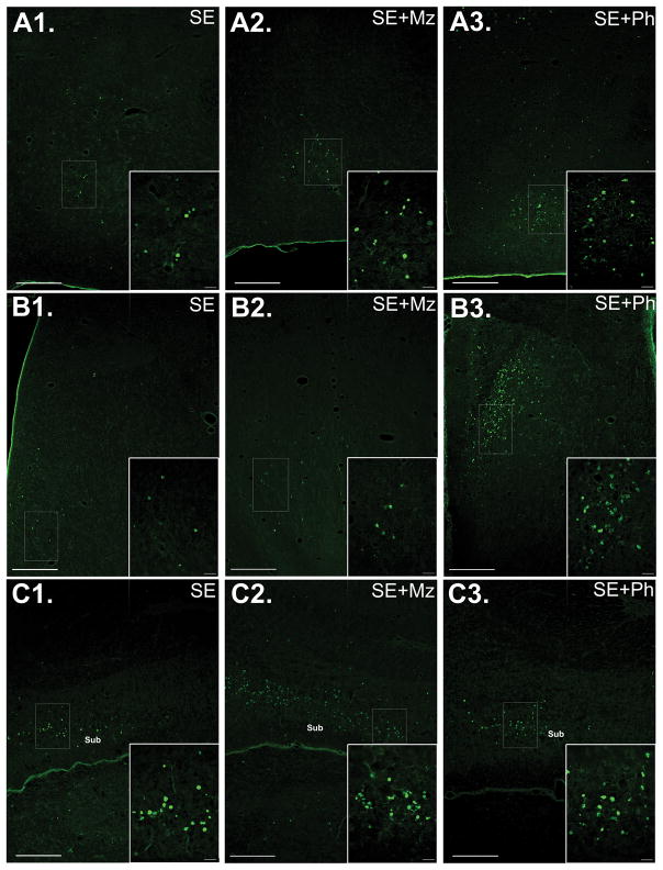
Figure 2. Midazolam or phenobarbital treatment increases SE-associated neuronal injury in some limbic regions.
(A) Images of FJB staining in hypothalamus of (A1) SE, (A2) SE+Mz, and (A3) SE+Ph pups, with a higher magnification of the boxed area on the bottom right of each image. Phenobarbital treatment significantly increased FJB+ cells compared to untreated SE. (B) Images of FJB staining in septal nuclei of (B1) SE, (B2) SE+Mz, and (B3) SE+Ph pups. Both midazolam and (to a greater extent) phenobarbital treatment significantly increase neuronal injury in septum. (C) Images of FJB staining in Ventral CA1/Subiculum of (C1) SE, (C2) SE+Mz, and (C3) SE+Ph P7 pups. As shown, SE (C1) induces neuronal injury in Ventral CA1/Subiculum by 24 hours post-SE and this injury is enhanced following midazolam treatment (C2), although no significant difference is seen after phenobarbital treatment (C3). Abbreviations: Sub=Subiculum. Scale bars: (A, B, C) long bars = 200 μm and short bars = 20 μm.

