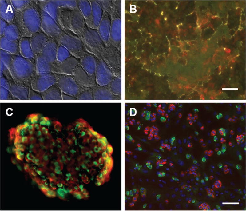
Figure 1.
Preparation of surface for islet cell culture system. (A) Human bladder carcinoma HTB-9 cells grown to confluence on 96- and 384-well plates. Overlay of bright-field and fluorescent Hoechst nuclear dye. (B) After denuding the cells from the plate, the plate surface was stained for fibronectin (red) and laminin (green) levels. Scale bar = 10 µm. (C) Immunofluorescence on an intact human islet, revealing cellular architecture and distribution of cells. Red, insulin; green, glucagon. (D) Immunofluorescence on dissociated islet cells seeded in a 384-well plate. Red, C-peptide; green, glucagon; blue, Hoechst nuclear dye. Scale bar = 100 µm.

