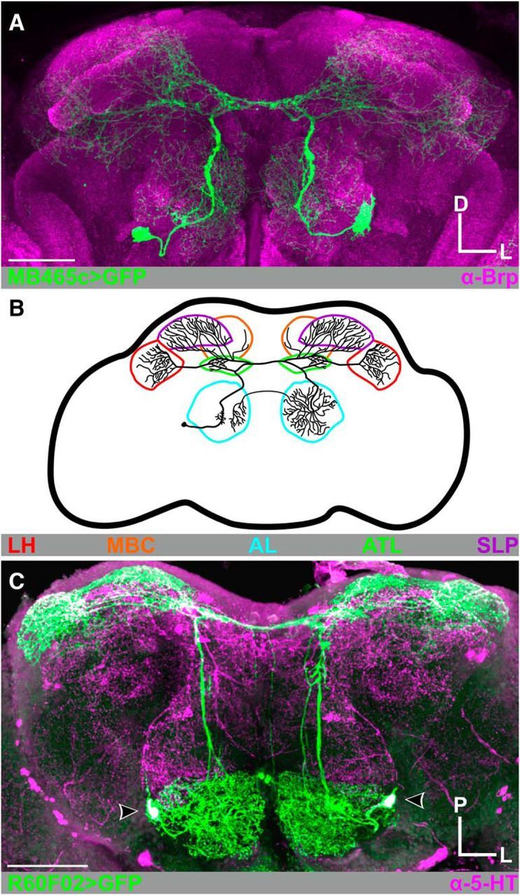
Figure 1.

CSDn morphology. A, MB465c-spGal4-driven expression of GFP (green) highlights the broadly projecting innervation pattern of the CSDns throughout the Drosophila brain. Neuropils are delineated with Brp-ir in magenta. B, Schematic of a single CSDn. The CSDns project throughout the olfactory system, including the antennal lobes (AL; cyan), antler (ATL; green), superior lateral protocerebrum (SLP; purple), mushroom body calyx (MBC; orange), and lateral horn (LH; red). In subsequent figures, diagrams represent specific regions of interest depicted in said figure. C, CSDns are 5-HT-immunoreactive (-ir). R60F02-Gal4-driven expression of GFP in the CSDns (green) completely overlaps with 5-HT-ir (magenta). Scale bars, 50 μm. D, Dorsal; L, lateral; P, posterior.
