Figure 3.

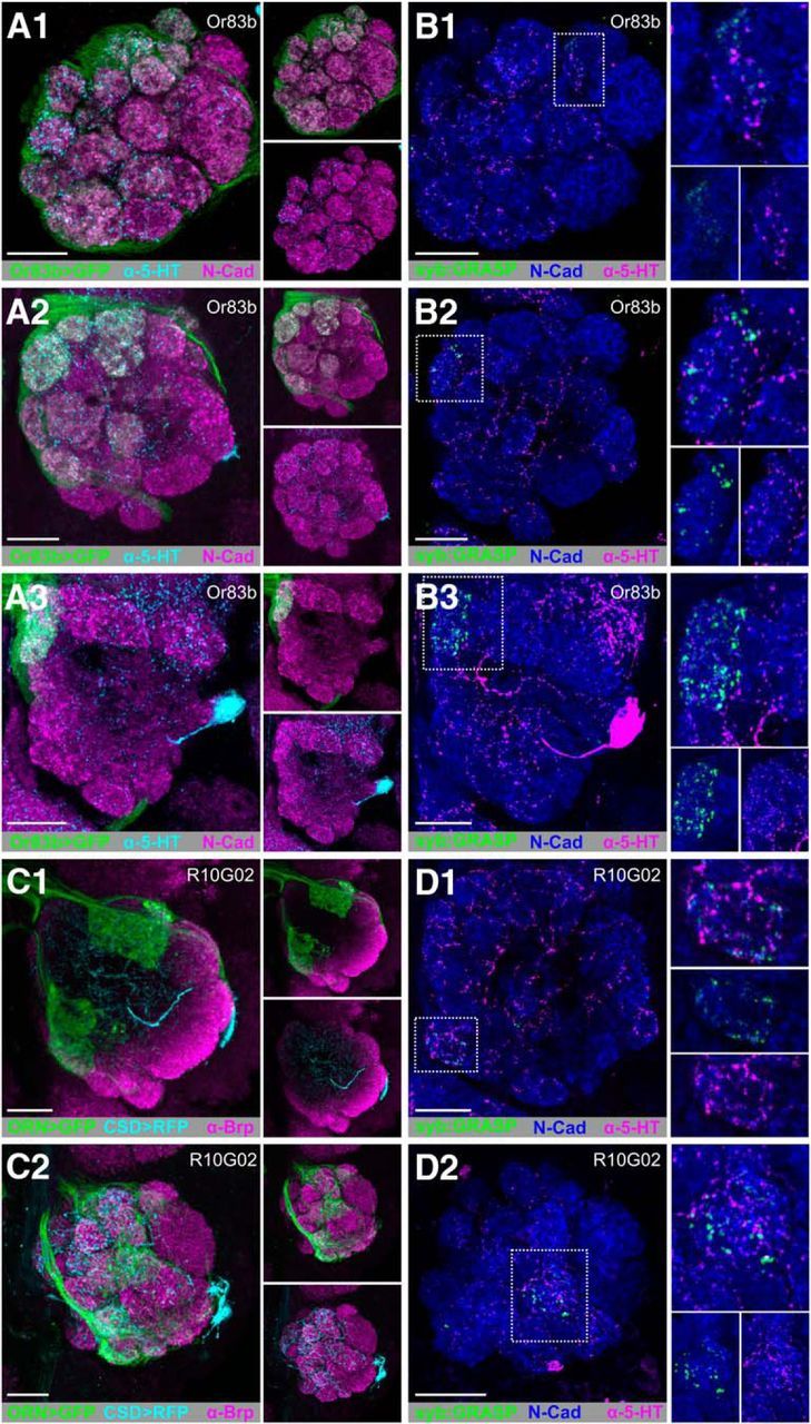
ORNs provide glomerulus-specific input to the CSDns in the AL. A, GFP-driven expression of Or83b-LexA (green) and 5HT-ir (cyan) in the AL at (A1) anterior, (A2) middle, and (A3) posterior depths. B, syb:GRASP signal of Or83b-LexA (presynaptic) and MB465c-spGal4 (postsynaptic) is located within DL4, DM2, and DM1 as depicted by scans at (B1) anterior, (B2) middle, and (B3) posterior depths. C, RFP (cyan) expression by the CSDns (via MB465c-spGal4) and GFP (green) expression by a subset of ORNs (via R10G02-LexA) in the AL. R10G02 labels a subset of ORNs, including some that express IRs. C1, Anterior, C2, Posterior. N-cadherin-ir delineates neuropil in A and B. Brp-ir delineates neuropil in C. D, syb:GRASP signal between R10G02-LexA (presynaptic) and MB465c-spGal4 (postsynaptic) is localized to just two glomeruli: VM1 (C1) and VA7l (C2). N-cadherin-ir delineates neuropil (blue). Reconstituted GFP (green) was identified as GFP-ir only in close association with 5-HT-ir processes (magenta). N-cadherin-ir delineates neuropil (blue) in D. syb:GRASP was observed less frequently in VA7l (B1) via R10G02 and DL4 (D2) via Or83b-LexA, suggesting interanimal variability. Scale bars, 20 μm.
