Figure 2.

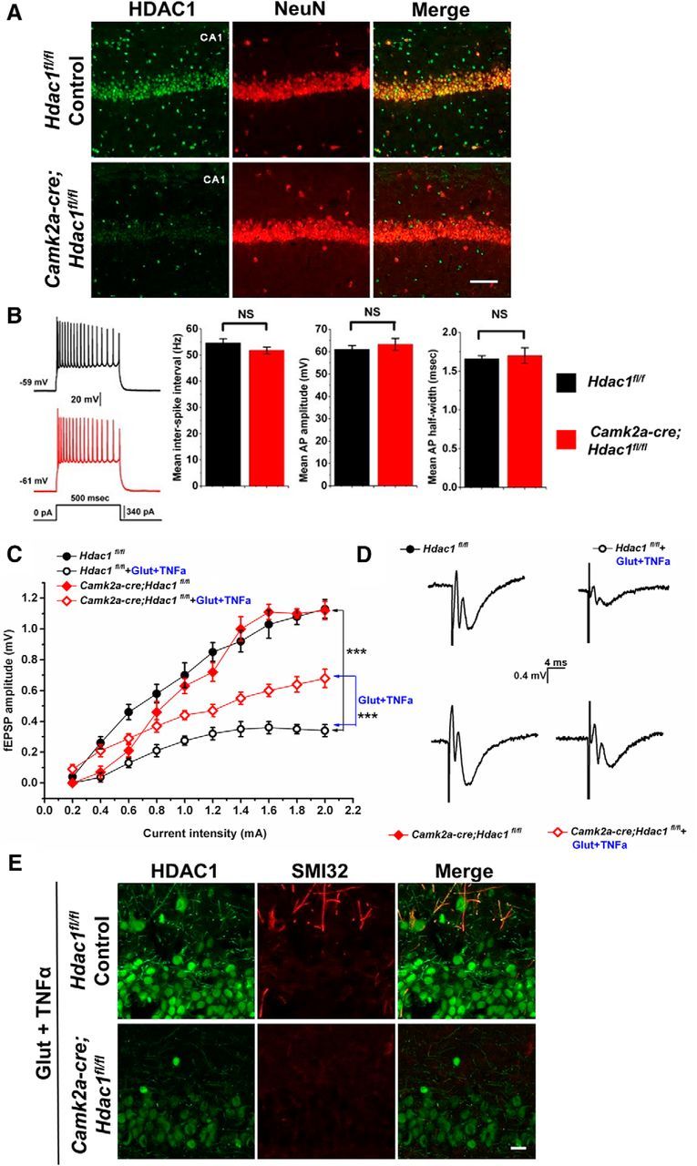
Conditional ablation of Hdac1 in Camk2a-cre;Hdac1fl/fl CA1 pyramidal neurons is protective from axonal damage caused by exposure to glutamate and TNFα. A, Micrographs through area CA1 from adult Hdac1fl/fl control (top row) or Camk2a-cre;Hdac1fl/fl conditional knock-out mice (bottom row) immunostained for HDAC1 (green) and NeuN (red). HDAC1 immunoreactivity is undetectable in CA1 pyramidal cells, but is still present in interneurons, as expected because such neurons do not express Camk2a. Scale bar, 100 μm. B, Representative examples of APs in Hdac1fl/fl control (black record; 9 cells from 4 animals) or Camk2a-cre;Hdac1fl/fl (red record; 7 cells from 4 animals) CA1 pyramidal neurons in response to depolarizing current injection. There were no significant differences in the mean interspike interval (p = 0.47), mean amplitude (p = 0.17), or mean half-width (p = 0.48) of APs in the floxed control CA1 neurons relative to the mutant CA1 neurons. Error bar indicates mean ± SEM. Data were analyzed with two-tailed Student's t tests. C, Comparison of I/O relationships across genotypes and experimental conditions. There were no differences in I/O relationships between untreated Hdac1fl/fl controls (black filled circles, n = 7 from 4 animals) or Camk2a-cre;Hdac1fl/fl mice (red filled diamonds, n = 7 from 4 animals). Floxed control slices challenged with 100 mm glutamate + 200 ng/ml TNFα for 1 h (black open circles, n = 5 from 4 animals) show a larger reduction in fEPSP amplitude compared with HDAC1 knock-out slices receiving the same glutamate/TNFα treatment (red filled diamonds, n = 7 from 4 animals) relative to baseline, suggesting that ablation of Hdac1 was partially protective of the damaging effects of Glut and TNFα on axonal and synaptic function. Data were analyzed with one-way ANOVA with Bonferroni post hoc test, **p = 0.0099, ***p = 7.9 × 10⁻¹¹. D, Representative fEPSP recordings from the four different conditions as indicated in C. E, Acute hippocampal slices treated with 100 μm glutamate and 200 ng/ml TNFα for 1 h were immunostained for HDAC1 (green) and nonphosphorylated neurofilament-H (SMI32, red) to assess axonal damage. Scale bar, 20 μm.
