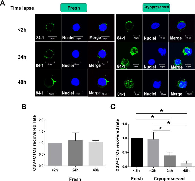
Figure 5. Comparison of CTCs counts within different time lapse before cryopreserving PBMCs.

(A) Micrographs showing cell surface vimentin staining in the fresh and cryopreserved samples with different time lapse before cryopreserving PBMCs: less than 2 h, 24h and 48h. Graph shows the results of CTCs counts among fresh sarcoma samples (B) and cryopreserved ones (C) with different time lapse before isolating PBMCs: less than 2h, 24h and 48h. CSV (84-1, green), nuclear stain (blue). GIST, Gastrointestinal stromal tumor. Scale indicates 10 μm. The group of time-lapse less than 2 hours was used as the reference.* P<0.05
