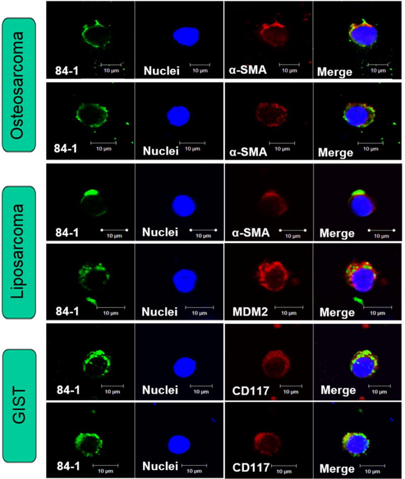
Figure 6. Independent tumor markers staining against captured CTCs using modified technique.

Captured CSV-positive CTCs validated by the presence of specific mesenchymal markers across 6 samples: α-SMA (red) for osteosarcoma, MDM2 (red) and α-SMA (red) for myxoid liposarcoma, and CD117 (red) for gastrointestinal stromal tumor (GIST). CSV (84-1, green), nuclear stain (blue). GIST, Gastrointestinal stromal tumor. Scale indicates 10 μm.
