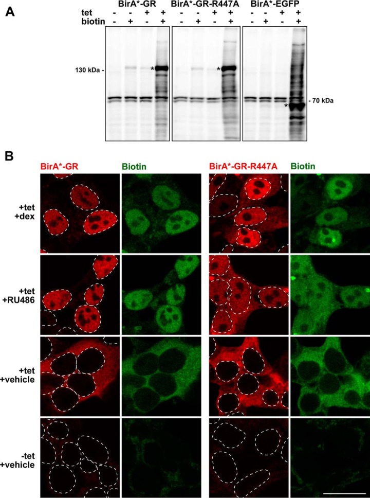
Fig. 1.
Validation of the cell lines expressing BirA*-fused GR, GR-R447A and EGFP. A, Streptavidin-HRP immunoblots of cells with (+) or without (-) added biotin (50 μm) and tetracycline (tet, 0.03 μg/ml). Asterisks depict fusion protein positions. B, Confocal fluorescence microscopy images of BirA*-GR and BirA*-GR-R447A-expressing cells treated with dexamethasone (dex, 100 nm), RU486 (1 μm) or vehicle in the presence of biotin and with or without tetracycline (tet, 0.03 μg/ml). BirA*-fusion proteins were detected with anti-HA (red) and biotinylated proteins with fluorescently labeled streptavidin (green). Dashed lines indicate positions of cell nuclei as inferred from DAPI DNA staining. Scale bar: 20 μm.

