
Figure 1.
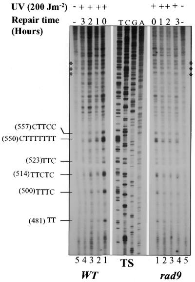

Repair of UVC-induced PDs in the GAL10 TS in G1-arrested wild-type and rad9 mutant cells. α-Factor-synchronized S.cerevisae cells were UV-irradiated and allowed to repair in the presence of α-factor for the indicated periods. NER was analyzed by primer extension. (A) Autoradiograms showing the primer extension products. Asterisks, non-specific Taq polymerase arrests; T, C, G and A, sequencing reactions. Each pyrimidine track on the left represent a PD cluster in the GAL10 gene with the accompanying number in parentheses referring to the 5′ nucleotide of the cluster. (B) Quantitative analysis of PD removal, illustrating the fraction (%) of PDs removed at each repair time. Each data point corresponds to an average value for the repair of several PD clusters. Inter-lane loading differences were corrected as described in the Materials and Methods. Each error bar represents the standard deviation of three experiments.