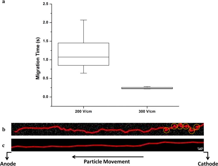
Figure 5.

(a) Box plot comparing the minimum, first quartile, median, third quartile, and maximum migration time (s) for polystyrene beads at 200 and 300 V/cm migrating throughout the entire length (100 μm) of a COC nanoslit. (b) Trace of a single PS bead translocating a 3 μm × 150 nm × 100 μm (w × d × l) channel under a field strength of 200 V/cm. Yellow circles indicate regions of possible recirculation. (c) Trace of a single PS bead translocating a 3 μm × 150 nm × 100 μm (w × d × l) channel under a field strength of 300 V/cm. The depth of focus of our 100× objective was large enough to ensure that each PS bead remained in focus since our channel depth was 150 nm.
