
Figure 2. Diminished low-theta power in lesioned PFC at active processing.
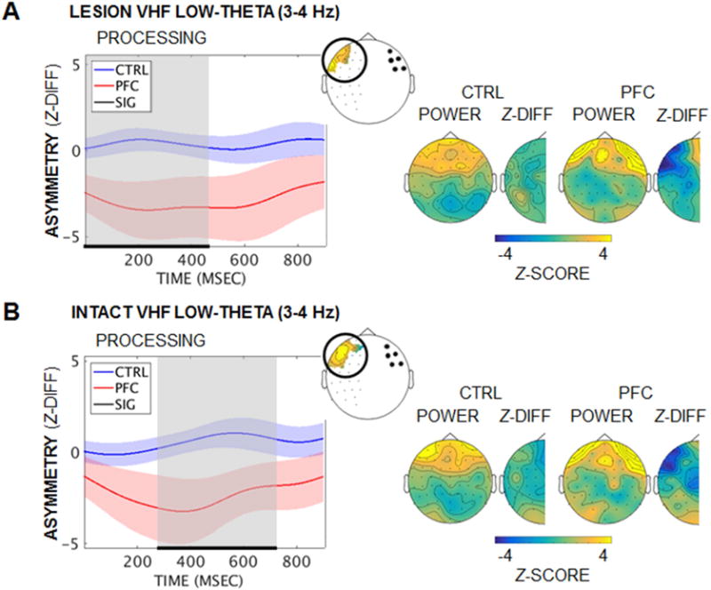
(A) Mean task-induced low-theta (3–4 Hz) hemispheric asymmetry in PFC over active processing by group when stimuli were presented to the lesioned visual hemifield. Low-theta power was diminished in patients in channels over the lesion, relative to the homologous intact-hemisphere channels (Group × Hemisphere pcluster = 0.04). Left panel: Significant effects are marked in black/gray and masked per channel on the BioSemi-64 topography (inset). Right panel: Scalp distributions of power and hemispheric difference z-scores are presented for the period of significant effects. While anterior theta power appears elevated in patients relative to controls, the contrast did not survive statistical testing (Group pcluster > 0.61). Shading = SEM; Z-DIFF, difference between lesioned- and intact-hemisphere z-scored power; VHF, visual hemifield. See also Figure S4.
(B) Equivalent to (A): Similar low-theta power effects were observed when stimuli were presented to the intact visual hemifield (Group × Hemisphere pcluster = 0.036).