
Figure 4. Nesprin1 is a widespread component of ciliary rootlets.
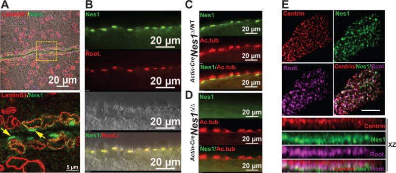
A. Nesprin1 and Lamin B1 immunolabeling of ependymal cells lining the lumen of wild-type brain ventricles. Note the bright staining of Nesprin1 arrays on the apical side of ependymal cells lining the ventricular lumen (arrows in inset). B. Colocalization of Nesprin1 with ciliary rootlets of ependymal cells labeled with rootletin C. Localization of Nesprin1 underneath ciliary axonemes of ependymal cells cilia labeled with acetylated tubulin (Ac.tub). D. Same experiments as in (C) performed on Actin-CreNes1Δ/Δ brains. Note the absence of Nesprin1 immunoreactivity at ciliary rootlets. E. Apical view of a single multiciliated MTEC colabeled with Centrin, Nesprin1 and Rootletin (top). Note the colocalization of rootletin and Nesprin1 just underneath the array of basal bodies labeled with Centrin in XZ projections (bottom).