
Figure 3. Silencing HoxA1 in human breast cancer cells reduces cell growth and restores lumen organization in tumor cell spheroid cultures.
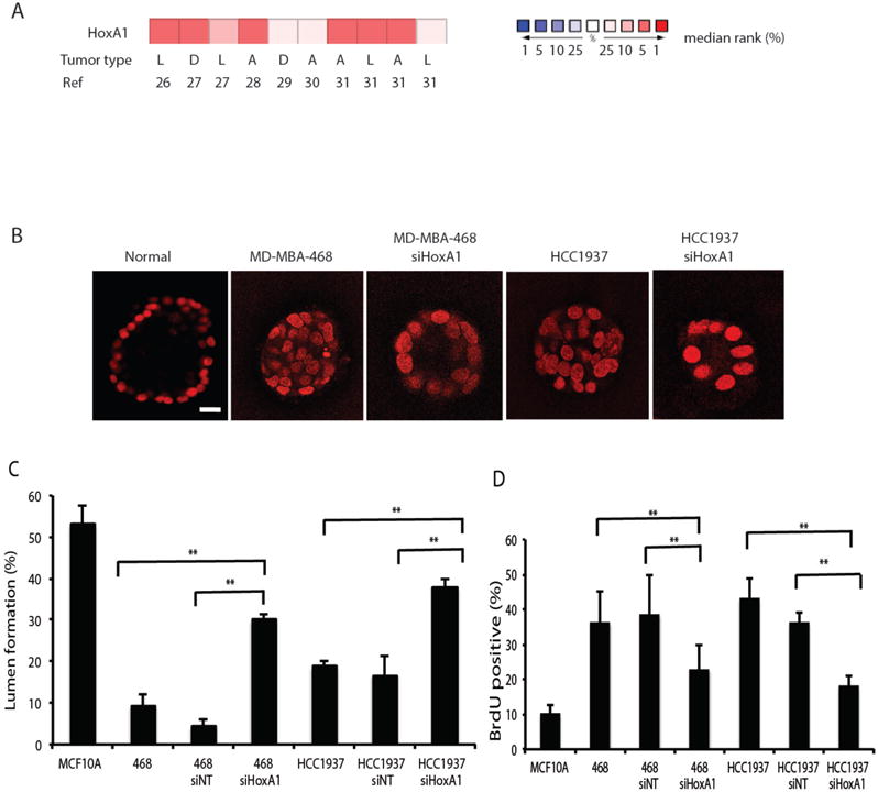
(A) The Oncomine compendium of cancer transcriptome profiles was used for analysis and visualization of HoxA1 expression data in human breast tumors and normal breast tissue. Differential expression analysis identified 10 patient datasets in which HoxA1 was over-expressed in human breast lesions by greater than 2-fold (see references 26–31). The type of tumor represented in the datasets is categorized as D, ductal breast carcinomas; L, lobular breast carcinomas; A; all breast carcinomas. The rank order of HoxA1 in each analysis is displayed (red, increased rank %; blue, decreased rank %). (B) Confocal images through the center of carmine-labeled spheroid cultures of normal human breast epithelial cells (MCF10A) and human breast cancer MD-MBA-468 and HCC1937 cell lines reveal. Breast cancer cells were also treated withsiHoxA1. Scale bar, 20 μm. (C) Hollow lumen formation and (D) proliferation of cancer cells in collagen-Matrigel spheroid cultures. Quantitation of lumen formation in non-tumorigenic human MCF10A breast epithelial cells and cells treated with scrambled siRNA controls are shown for comparison. Data are means +/− SD (n = 3, 150–300 cells per sample) **p<0.01, Fisher’s exact t-test.