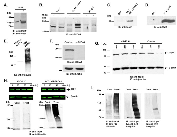
Figure 3. BRCA1 associates and ubiquitinates topoI.

A. Control and CPT-treated (5 μM SN-38) HCT-15 cells were lysed and subjected to immunoprecipitation with anti-BRCA1. Immunoprecipitates were immunoblotted with anti-topoI. B. Control and CPT-treated (5 μM SN-38) HCT-15 cell lysates were subjected to immunoprecipitation with anti-topoI or IgG (control) and immunoprecipitates were immunoblotted with anti-BRCA1. C. GST and GST-BRCA1-BRCT were incubated with HCT-15 cell lysate. After extensive washing the adsorbates were analyzed by immunoblotting with anti-topoI. D. GST and GST- topoI (aa1-210) were incubated with HCT-15 cell lysates. After extensive wash the adsorbates were analyzed by immunoblot analysis with anti-BRCA1. E. GST-topoI was phosphorylated with DNA-PK and then incubated with purified E1, UbcH5c (E2), and BRCA1/BARD1 (E3) and without BRCA1/BARD1. The reaction products were immunoblotted with anti-ubiquitin. F. BT-474 cells were transduced with BRCA1 silencing lentivirus (shBRCA1) and scrambled sequence lentivirus (as control). Control and shBRCA1 cells lysates were analyzed by immunoblotting with anti-BRCA1 and anti-β-actin. G. Control and shBRCA1 BT-474 cells were treated with 5 μM SN-38 for 2h and 6h and the cell lysates were immunoblotted with anti-topoI and anti-β-actin. H. HCC1937 and HCC1937-BRCA1, cells were treated with 5 μM SN-38 for 30, 60 and 90 min. Cell lysates were immunoblotted with anti-topoI (upper panel) and β-actin (middle panel). Cell lysates were also subjected to immunoprecipitation with anti-topoI and immunoprecipitates were immunoblotted with anti-ubiquitin (lower panel). I. HCC1937-BRCA1 cells were treated with 5 μM SN-38 and cell lysates were subjected to immunoprecipitation with anti-topoI. The immunoprecipitates were analyzed by immunoblot with the indicated antibodies.
